Jing-yan Liu1,2,
Yu-juan Guo2,
Yong-zhan Song3,
Xing-liang Song4,
Dian-jie Liu1 ![]()
For correspondence:- Dian-jie Liu Email: lindjsd@126.com Tel:+8653187938911
Received: 30 September 2016 Accepted: 21 December 2016 Published: 31 January 2017
Citation: Liu J, Guo Y, Song Y, Song X, Liu D. Expressions of Wingless and Int1 (Wnt)-induced secreted protein 1 in paraquat-poisoned patients. Trop J Pharm Res 2017; 16(1):225-230 doi: 10.4314/tjpr.v16i1.30
© 2017 The authors.
This is an Open Access article that uses a funding model which does not charge readers or their institutions for access and distributed under the terms of the Creative Commons Attribution License (http://creativecommons.org/licenses/by/4.0) and the Budapest Open Access Initiative (http://www.budapestopenaccessinitiative.org/read), which permit unrestricted use, distribution, and reproduction in any medium, provided the original work is properly credited..
Purpose: To study the ex
Methods: A total of 37 PQ-poisoned patients were enrolled in the study, and divided into non-survivor group (NS) and survival group (S) based on the final therapeutic outcome. Besides, another normal control group (NC) comprised of normal healthy people. Serum PQ concentration was determined by high performance liquid chromatography (HPLC), while reverse transcription-polymerase chain reaction (RT-PCR) and enzyme-linked immunosorbent assay (ELISA) were used to evaluate WISP1 in the serum of PQ poisoned patients.
Results: PQ intake in NS and S groups were 23.58 ± 26.23 and 143.18 ± 263.04 mL, respectively, while serum PQ concentration was 2.07 ± 0.67 and 4.12 ± 1.74 mg/L, respectively. Significant correlation was found between the outcome of patients and serum PQ concentration (OR = 1.434, p < 0.01). Serum PQ concentration was closely correlated with WISP1 gene ex
Conclusion: WISP1 is over-expressed in PQ-poisoned patients, and serum PQ concentration may be a useful index for the prognosis of PQ poisoned patients.
Introduction
Paraquat (PQ), also named as 1,1-dimethyl-4,4-bipyridinium dichloride, is commonly used as a non-selective herbicide. However, PQ can result in acute poisoning via intake through digestive tract and mucous membrane. Currently, it’s reported that PQ poisoning has a very high morbidity ranging from 30.0 to 88.89 % [1-3]. PQ could lead to the over-releases of many cytokines in PQ poisoned animal and humans. Wingless & Int1 (Wnt)-induced secreted protein 1 (WISP1), a matricellular protein of CCN family, regulates proliferation, survival and migration of cells. Furthermore, WISP1 plays an important effect on wound repair, tumorigenesis and angiogenesis [4-6]. It is reported that WISP1 is a mediator of disturbed epithelial-mesenchymal crosstalk. WISP1 could be up-regulated in experimental lung fibrosis and could promote the development of pulmonary fibrosis in vivo [7]. WISP1 was also up-regulated in alveolar epithelial type II cells with stretch-induced epithelial-mesenchymal transition (EMT) in vitro [8], and related to ventilator-induced lung injury models in vivo [9].
The previous reports indicate that WISP1 plays a crucial role in pulmonary injury and fibrosis. Thus, we hypothesize that WISP1 may play an important role in the development of pulmonary injury and fibrosis induced by PQ. However, expression changes of WISP1 in PQ poisoned patients have not been reported so far. This study was designed to investigate WISP1 expression in serum of PQ-poisoned patients and explore the relationship between PQ concentration and WISP1 levels in PQ-poisoned patients.
Methods
Subjects
The 20 % PQ (w/v) poisoned patients were collected in Linyi Peoples’s Hospital (Linyi, China) from Jan. 2013 to Jun. 2014. The PQ intake ranges of these patients were from one mouthful to 20 mL. The patients were divided into non-survivor (NS) and survivor (S) groups according to the final therapeutic outcome. Besides, another normal control group (NC) was also set by normal healthy volunteers. Twenty female and 17 male PQ-poisoned patients with a mean age of 30 years (ranging from 20 to 59 years old) were enrolled in the study. The protocols of this study were approved by the Ethics Committee of the Linyi Peoples’s Hospital (approval no. 2013-J-03) and in accordance with the Helsinki declaration [10]. The written consent of all subjects and their relatives was obtained.
Blood sample collection
Venous blood samples (6 mL) were collected in vacuum blood collection tube from each PQ-poisoned patient on the first and third days in the hospital. Healthy control participants’ venous blood samples were collected in the morning. The blood samples were centrifuged (3000 g, for 10 min) and the serum samples were collected and kept at -80 °C until assays.
Determination of PQ in serum
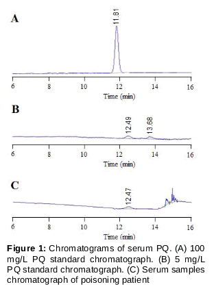
An Agilent Zorbax HC-C18 (2.1 mm × 150 mm, 5 μm) column was applied for chromatographic analysis with temperature set at 30 °C. The flow-rate of the mobile phase A (acetonitrile, 15 %) was set as 1.0 mL/min. Mobile phase B contained 260 mM sodium dihydrogen phosphate and 20mM sodium heptanesulfonate. The pH was adjusted to 2.0 with triethylamine. The detection wavelength was 256 nm for PQ.
Sample preparation
A 400 μL aliquot of patient’s serum was added to 500 μL of acetonitrile, and the mixture was mixed for 30 s in a vortex mixer. Then, the mixed sample was centrifuged (7000 g, for 5 min) to obtain the supernatants. After that, the supernatants were mixed with 3 mL trichloromethane (CHCl3) and then mixed. Subsequently, the mixture was again centrifuged (7000 g, for 5 min). The lower colorless transparent liquid (20 μL) was collected for the tests.
Calibration curve
Six calibration serum standards (0.5, 1.0, 2.5, 5.0, 10.0, and 100.0 μg/mL) were prepared by spiking 400 μL blank human serum with 600 μL of the working solutions of PQ. After internal standard solution was added and deproteinization was performed, the serum mixture was introduced into the HPLC column, and PQ levels were measured by the internal standard method.
Total RNA extraction and cDNA preparation
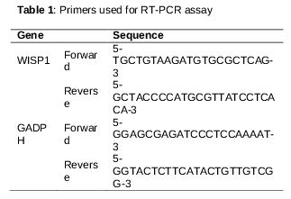
Total RNA was extracted using Trizol reagent (Sigma Alorich Co. LLC), and the RNA concentration was quantitated by spectrophotometry and agarose gel electrophoresis. The mixture of 1 µL of total RNA and 13 μL water was incubated at 65 °C for 5 min. Then successively add 4 μL 5 × M-MLV buffer, 1 μL of M-MLV RTase and 1 µL of primer. The mixture was incubated at 37 °C for 15 min, and reverse transcriptase was performed by heating at 98 °C for 5 min. Then, the cDNA was kept at -20 °C. Primer sequences for WISP-1 were obtained according to previous study [7]. Primers of GAPDH gene were designed according to the online ensemble database (http://www.ensembl.org). Primers used in this research were purchased from Invitrogen Life Co., Shanghai, China ().
Reverse transcription-polymerase chain reaction (RT-PCR) assay
Conditions for reverse transcription-polymerase chain reaction (RT-PCR) assays were optimized by using iCycler iQ (BioRad, USA). Hot start PCR was performed using a program of 5 min denaturation at 95°C, followed by 35 cycles (95 °C for 30 s, 55 °C for 20 s, and 72 °C for 30 s), and then a final 10 min extension at 72 °C. Then 2 μL cDNA was analyzed on 1.5 % agarose gel. Gel imaging system (Universal Hood II) was used to take UV photography. The gel photograph result was analyzed by Gel-Pro Analyzer Version 3.0 gel image analysis system.
Enzyme-linked immunosorbent assay (ELISA)
WISP1 protein levels in the blood serum were determined by using commercial WISP-1 (CCN4) human enzyme-linked immunosorbent assay (ELISA) kit (Abcam, USA) according to the instructions described by the manufacturer.
Statistical analysis
Data were analyzed by SPSS 19.0 statistical software, and are presented as mean ± standard deviation (SD). The t-test was used to investigate the differences among quantitative variables. The relationships between categorical variables and outcomes were evaluated using Spearman correlation, Chi square test and Logistics regression. In all cases, a confidence interval of 95 % and a level of p < 0.05) were considered significant.
Results
Demography and mortality
There were 37 patients (20 females and 17 males) collected in the study. The age and PQ intake amount of PQ poisoned patients’ were 30.97 ± 11.54 years and 59.34 ± 150.90 mL, respectively. There were 11 patients in the non-survivor group (NS) and 26 patients in the survival group (S). There were 24 PQ-poisoned patients with multi-organ failure, especially acute kidney injury. Besides, 13 patients had acute lung injury and 9 patients died from acute respiratory failure.
Serum PQ concentration
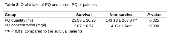
As shown in , the intake amount of PQ of the S and NS groups were 23.58 ± 26.23 mL and 143.18 ± 263.04 mL, respectively. Furthermore, the serum contents of PQ were determined by HPLC assay (). The results were showed in indicated that the serum contents of PQ of the S and NS patients were 2.07 ± 0.67 mg/L and 4.12 ± 1.74 mg/L, respectively, which also represented an obvious difference (p < 0.01). There was no obvious relationship between PQ intake amount and serum PQ contents (RR = 0.078, p = 0.647).
mRNA expressions of serum WISP1 genes
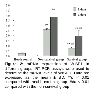
As can be seen from the , mRNA expressions of WISP1 in serum were presented. The mRNA expressions of WISP1 in non-survival and survival groups at the first poisoned day were higher than those of health group (p < 0.01, p < 0.01) and mRNA expressions of WISP1 in survival group in the 3rd day were down-regulated compared with the NS group (p < 0.01, p < 0.01).
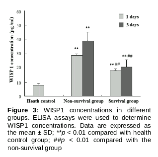
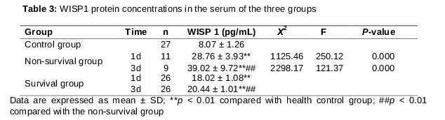
Serum WISP1 protein level
WISP1 serum concentrations are presented in . WISP1 protein serum concentration of the non-survivor group on the first day of poisoning was higher than those of health control group (p < 0.01) and the survival group (p < 0.01). Similar to the first day, WISP1 protein serum levels of the non-survivor group on the third day of poisoning day were also higher than those of healthy control (p < 0.01) and survival group (p < 0.01). Besides, WISP1 protein levels in survival group at the first poisoned day and (p < 0.01) third poisoned day (p < 0.01) were higher than that in health control group.
Relationship between serum PQ concentration and WISP1 expression
As can be seen from , the results of correlation analysis showed that serum PQ contents was closely correlated with the WISP1 gene expression on the first day of poisoning (OR = 0.621, p < 0.01). A correlation between the serum PQ and the serum WISP1 concentrations also occurred on the first (OR = 0.596, p < 0.01) and third days of poisoning (OR = 0.447, p < 0.01). No obvious relationship was found between serum PQ levels and WISP1 gene expression (OR = 0.164, p > 0.05) on the third day of poisoning.
Serum PQ concentration and prognosis
Further, the relationship between PQ concentrations and prognosis of PQ intake patients was also analyzed by logistic regression analysis. The results showed that a higher serum PQ concentration was closely correlated to higher mortality of PQ-poisoned patients (OR = 1.434, p < 0.05).
Discussion
PQ poisoning often result in death within a few days due to multiple organ failure (MOF), especially kidney and lung which are the target organs most affected. It has been reported that most of the survivors suffer from complicated pulmonary fibrosis (PF). In the study, 37 PQ poisoned patients were investigated, and the mortality rate was 29.73 %. The early symptom of PQ poisoning is alveolitis, as the lungs show alveolar collapse and inflammatory cells infiltrate into alveolar airspaces [11,12]. A previous report using 99mTc diethylenetriamine pentaacetate (DTPA) radioaerosol lung scintigraphy revealed that alveolar permeability is increased in PQ-poisoned patients [13]. Lung alveolitis reduces lung volume and lung compliance, and in the late stage of PQ intoxication, pulmonary fibrosis and respiratory failure is induced, finally leading to the death of PQ-poisoned patients. In this study, nine PQ poisoned patients were finally died from acute respiratory failure.
It is estimated that the lethal dose of PQ in an adult human is about 30 mg/kg [14,15] or 3 - 6 g of PQ ion [16,17]. A previous report showed that ingestion of > 30 mL may lead to a fatal outcome in PQ-poisoned patients [18]. It was observed that patients with higher serum PQ concentration led to higher mortality.
WISP1 is a secreted matricellular protein, which belongs to the CCN family. In humans, WISP1 is expressed in various organs, including the lung, heart, pancreas, kidney, placenta, small intestine, and spleen, etc [19]. Changes in WISP1 expressions in PQ-poisoned patients remain unknown. It is reported that PQ can induce increase in TGF-β1 and TNF-α in PQ poisoned patients and animals [20-24]. Long-term low-dose PQ exposure can induce EMT-like cellular transformation and subsequent fibrogenesis in A549 and human bronchial epithelial cells. It has also been reported that TGF-β1 plays an important role in EMT-like cellular response and subsequent fibrogenesis of PQ induced pulmonary cells [25]. WISP1 was increased in alveolar epithelial type II cells with stretch-induced EMT [8]. TGF-β1 and TNF-α induced WISP1 in primary human lung fibroblasts, WISP1 exerts its profibrotic effects through IL-6-dependent induction of fibroblast proliferation [26]. The results revealed that both mRNA and protein levels of WISP1 were highly elevated in PQ-poisoned patients, and that the expressions and serum contents of WISP1 in non-surviving patients were higher than those of surviving patients. In addition, serum PQ concentration was closely correlated with expression and serum concentration of WISP1 in PQ-poisoned patients.
Conclusion
WISP1 is up-regulated in PQ-poisoned patients, and serum PQ concentration is closely correlated with WISP1 expression. Furthermore, determina-tion of serum PQ concentration and WISP 1 would be beneficial for forecasting the prognosis of PQ poisoned patients.
Declarations
Acknowledgement
References
Archives


News Updates